Publication
666
Anal. Chem., 82 (15), 6353–6362, 2010.
(Accelerated Article)
DOI: 10.1021/ac1012464
|
|
|
|
|
 |
Touching Surface-Attached Molecules with a Microelectrode: Mapping the Distribution of Redox-Labeled Macromolecules by Electrochemical-Atomic Force Microscopy |
|
|
|
|
Agnès Anne, Edmond Cambril, Arnaud Chovin and Christophe Demaille
Laboratoire d’Electrochimie Moléculaire, Unité Mixte de Recherche Université CNRS No. 7591, Université Paris Diderot-Paris 7, 15 Rue Jean-Antoine de Baïf, 75205 Paris Cedex 13, France, and Laboratoire de Photonique et de Nanostructures (LPN/CNRS), Route de Nozay, 91460 Marcoussis, France
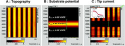
We report on the development of a mediator-free electrochemical-atomic force microscopy (AFM-SECM) technique designed for high-resolution imaging of molecular layers of nanometer-sized redox-labeled (macro)molecules immobilized onto electrode surfaces. This new AFM-SECM imaging technique, we call molecule touching atomic force electrochemical microscopy (Mt/AFM-SECM), is based on the direct contact between surface-anchored molecules and an incoming microelectrode (tip). To validate the working-principle of this microscopy, we consider a model system consisting of a monolayer of nanometer long, flexible, polyethylene glycol (PEG) chains covalently attached by one extremity to a gold surface and bearing at their free end a ferrocene (Fc) redox tag. Using Mt/AFM-SECM in tapping mode, i.e., by oscillating the tip so that it comes in intermittent contact with the grafted chains, we show that the substrate topography and the distribution of the redox-tagged PEG chains immobilized on the gold surface can be simultaneously and independently imaged at the sub-100 nm scale. This novel type of SECM imaging may be found useful for characterizing the surface of advanced biosensors which use electrode-grafted, redox-tagged, linear biochains, such as peptides or DNA chains, as sensing elements. In principle, Mt/AFM-SECM should also permit in situ imaging of the distribution of any kind of macromolecules immobilized on electrode surfaces or simply conducting surfaces, provided they are labeled by a suitable redox tag. |