Publication
692
Anal. Chem. 83 (20), 7924-7932, 2011
DOI: 10.1021/ac201907v
|
|
|
|
|
 |
High-Resolution Mapping of Redox-Immunomarked Proteins Using Electrochemical–Atomic Force Microscopy in Molecule Touching Mode |
|
|
|
|
Agnès Anne, Arnaud Chovin, Christophe Demaille, and Manon Lafouresse
Laboratoire d’Electrochimie Moléculaire, UMR 7591 CNRS, Université Paris Diderot, Sorbonne Paris Cité, 15 rue Jean-Antoine de Baïf, F-75205 Paris Cedex 13, France
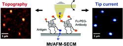
We explore the possibility of using molecule touching atomic force electrochemical microcopy (Mt/AFM–SECM) for high-resolution mapping of proteins on conducting surfaces. The proposed imaging strategy relies on making surface-immobilized proteins electrochemically “visible” via redox-immunomarking by specific antibodies conjugated to poly(ethylene glycol) (PEG) chains terminated by redox ferrocene (Fc) heads. The flexibility and length of the PEG chains are such that, upon approaching a combined AFM–SECM microelectrode tip toward the surface, the Fc moieties can efficiently shuttle electrons from the surface to the tip. The so-generated SECM positive feedback tip current allows the specific localized detection of the sought protein molecules on the surface. This new electrochemical imaging scheme is validated experimentally on the basis of a model system consisting of mouse IgGs adsorbed onto electrode surfaces and recognized by Fc–PEG-labeled antimouse antibodies. In order to estimate the resolution of Mt/AFM–SECM for protein imaging, regular arrays of submicrometer-sized spots of mouse IgGs are fabricated onto gold electrode surfaces using particle lithography. The Fc–PEG-immunomarked mouse IgG spots are imaged by Mt/AFM–SECM operated in tapping mode. Both an electrochemical image, reflecting the surface distribution of the redox-labeled IgGs, and a topography image are then simultaneously and independently acquired, with a demonstrated resolution in the 100 nm range. The strength of Mt/AFM–SECM imaging is to combine the nanometric resolution of AFM with the selectivity of the electrochemical detection, potentially allowing individual target proteins to be identified amidst similarly sized “nano objects” present on a conducting surface. |