Publication
773
ACS Nano, 9 (5),4911-4924, 2015
DOI:10.1021/acsnano.5b00952
|
|
|
|
|
|
 |
Electrochemical Atomic Force Microscopy Imaging of Redox-Immunomarked Proteins on Native Potyviruses: From Subparticle to Single-Protein Resolution |
|
|
|
Laurent Nault, Cécilia Taofifenua, Agnès Anne, Arnaud Chovin, Christophe Demaille, Jane Besong-Ndika, Daniela Cardinale, Noëlle Carette, Thierry Michon, and Jocelyne Walter
Laboratoire d’Electrochimie Moléculaire, Université Paris Diderot, Sorbonne Paris Cité, Unité Mixte de Recherche Université, CNRS No 7591, Bâtiment Lavoisier, 15 rue Jean-Antoine de Baïf, 75205 Cedex 13 Paris, France
UMR 1332 Biologie du Fruit et Pathologie, INRA-Université Bordeaux 2, 71 av. Edouard Bourlaux, 20032-33882 Cedex Villenave d’Ornon, France
Department of Food and Environmental Sciences, University of Helsinki, Latokartanonkaari 11, FI-00014 Helsinki, Finland
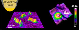
We show herein that electrochemical atomic force microscopy (AFM-SECM), operated in molecule touching (Mt) mode and combined with redox immunomarking, enables the in situ mapping of the distribution of proteins on individual virus particles and makes localization of individual viral proteins possible. Acquisition of a topography image allows isolated virus particles to be identified and structurally characterized, while simultaneous acquisition of a current image allows the sought after protein, marked by redox antibodies, to be selectively located. We concomitantly show that Mt/AFM-SECM, due to its single-particle resolution, can also uniquely reveal the way redox functionalization endowed to viral particles is distributed both statistically among the viruses and spatially over individual virus particles. This possibility makes Mt/AFM-SECM a unique tool for viral nanotechnology. |