Publication
781
Inorg. Chem., 54 (19), 9470-9482, 2015
DOI:10.1021/acs.inorgchem.5b01330
|
|
|
|
|
|
 |
De novo design and characterization of copper metallopeptides inspired by native cupredoxins |
|
|
|
Jefferson S. Plegaria, Matteo Duca, Cédric Tard, Thomas J. Friedlander, Aniruddha Deb, James E. Penner-Hahn, and Vincent L. Pecoraro
Department of Chemistry and Biophysics Department, University of Michigan, Ann Arbor, Michigan 48109, United States
Laboratoire d’Electrochimie Moléculaire, UMR 7591, CNRS, Université Paris Diderot, Sorbonne Paris Cité, 15 Rue Jean Antoine de Baïf, F-75205 Paris Cedex 13, France
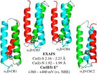
Using de novo protein design, we incorporated a copper metal binding site within the three-helix bundle a3D (Walsh et al. Proc. Natl. Acad. Sci. U.S.A. 1999, 96, 5486–5491) to assess whether a cupredoxin center within an a-helical domain could mimic the spectroscopic, structural, and redox features of native type-1 copper (CuT1) proteins. We aimed to determine whether a CuT1 center could be realized in a markedly different scaffold rather than the native ß-barrel fold and whether the characteristic short Cu–S bond (2.1–2.2 Å) and positive reduction potentials could be decoupled from the spectroscopic properties (e600 nm = 5000 M–1 cm–1) of such centers. We incorporated 2HisCys(Met) residues in three distinct a3D designs designated core (CR), chelate (CH), and chelate-core (ChC). XAS analysis revealed a coordination environment similar to reduced CuT1 proteins, producing Cu–S(Cys) bonds ranging from 2.16 to 2.23 Å and Cu–N(His) bond distances of 1.92–1.99 Å. However, Cu(II) binding to the CR and CH constructs resulted in tetragonal type-2 copper-like species, displaying an intense absorption band between 380 and 400 nm (>1500 M–1 cm–1) and A|| values of (150–185) × 10–4 cm–4. The ChC construct, which possesses a metal-binding site deeper in its helical bundle, yielded a CuT1-like brown copper species, with two absorption bands at 401 (4429 M–1 cm–1) and 499 (2020 M–1 cm–1) nm and an A|| value ~30 × 10–4 cm–4 greater than its native counterparts. Electrochemical studies demonstrated reduction potentials of +360 to +460 mV (vs NHE), which are within the observed range for azurin and plastocyanin. These observations showed that the designed metal binding sites lacked the necessary rigidity to enforce the appropriate structural constraints for a Cu(II) chromophore (EPR and UV–vis); however, the Cu(I) structural environment and the high positive potential of CuT1 centers were recapitulated within the a-helical bundle of a3D. |